1. Structure and Function of the Nervous System
The Central and Peripheral Nervous Systems
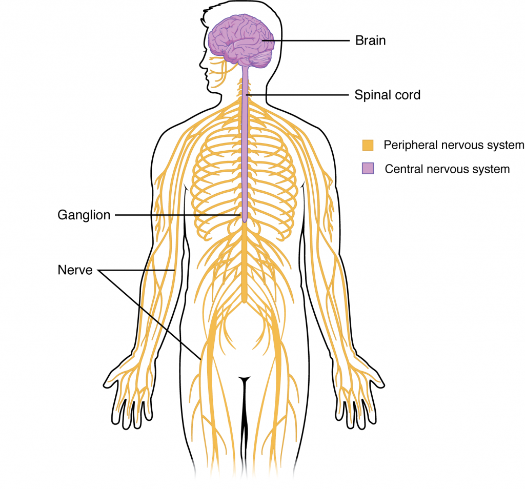
The picture you have in your mind of the nervous system probably includes the brain, the nervous tissue contained within the cranium, and the spinal cord, the extension of nervous tissue within the vertebral column. Additionally, the nervous tissue that reach out from the brain and spinal cord to the rest of the body (nerves) are also part of the nervous system. We can anatomically divide the nervous system into two major regions: the central nervous system (CNS) is the brain and spinal cord, the peripheral nervous system (PNS) is the nerves (Figure 1.1). The brain is contained within the cranial cavity of the skull, and the spinal cord is contained within the vertebral canal of the vertebral column. The peripheral nervous system is so named because it is in the periphery—meaning beyond the brain and spinal cord.
Functional Divisions of the Nervous System
In addition to the anatomical divisions listed above, the nervous system can also be divided on the basis of its functions. The nervous system is involved in receiving information about the environment around us (sensory functions, sensation) and generating responses to that information (motor functions, responses) and coordinating the two (integration).
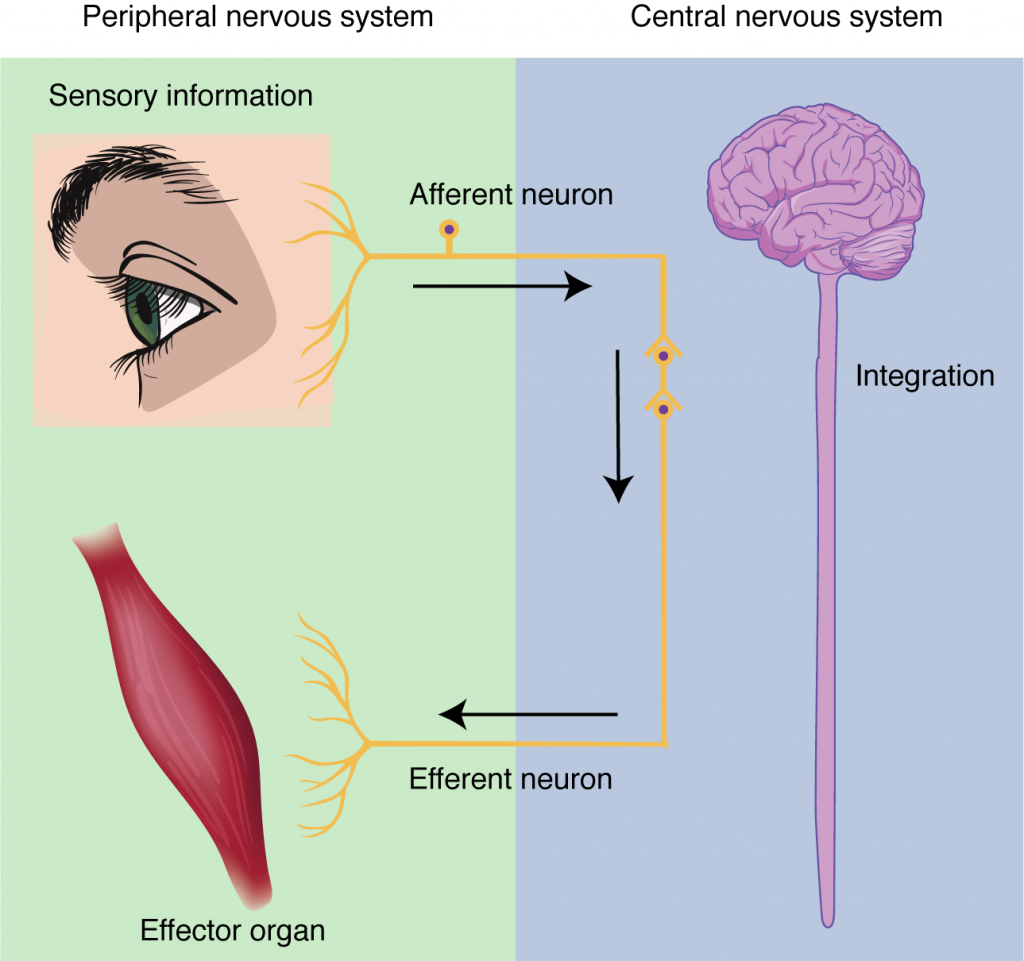
Sensation. Sensation refers to receiving information about the environment, either what is happening outside (ie: heat from the sun) or inside the body (ie: heat from muscle activity). These sensations are known as stimuli (singular = stimulus) and different sensory receptors are responsible for detecting different stimuli. Sensory information travels towards the CNS through the PNS nerves in the specific division known as the afferent (sensory) branch of the PNS. When information arises from sensory receptors in the skin, skeletal muscles, or joints, it is transmitted to the CNS using somatic sensory neurons; when information arises from sensory receptors in the blood vessels or internal organs, it is transmitted to the CNS using visceral sensory neurons.
Response. The nervous system produces a response in effector organs (such as muscles or glands) due to the sensory stimuli. The motor (efferent) branch of the PNS carries signals away from the CNS to the effector organs. When the effector organ is a skeletal muscle, the neuron carrying the information is called a somatic motor neuron; when the effector organ is cardiac or smooth muscle or glandular tissue, the neuron carrying the information is called an autonomic motor neuron. Voluntary responses are governed by somatic motor neurons and involuntary responses are governed by the autonomic motor neurons, which are discussed in the next section.
Integration. Stimuli that are detected by sensory structures are communicated to the nervous system where information is processed. In the CNS, information from some stimuli is compared with, or integrated with, information from other stimuli or memories of previous stimuli. Then, a motor neuron is activated to initiate a response from the effector organ. This process during which sensory information is processed and a motor response generated is called integration (see Figure 1.2 below).